

Coexistence of Onychomycosis and Tinea Pedis in Patients with Diabetes
- Ektaa Vadgama

- Aug 30, 2021
- 4 min read

I was fortunate enough to be placed in a clinic that saw high-risk patients with acute wounds. It was a great learning opportunity for me but it was glaringly obvious that diabetic patients had a couple of things going on in their feet. So after looking for some articles to use as a reference in my placement reflections, I came across one particular article discussing the prevalence of diabetic patients with onychomycosis and tinea pedis.
Coexistence of Onychomycosis and Tinea Pedis in Patients with Diabetes
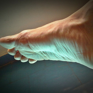
Diabetes is a condition that influences the progression or introduction of new complications to the body (Rich, et al., 2003). The most problematic of the complications being lower limb ulcerations that can result in amputation thereby reducing the patient’s life span and overall quality of life (Jacobson, et al., 1994). With most patient presentations that were seen by myself in clinic today, a majority leaned towards onychomycosis and tinea pedis, often presenting together, which begs the question, why?
In order to answer that question, it’s easier to break down the epidemiology and pathophysiology of cutaneous fungal infections and look at the clinical evidence and associations of onychomycosis amongst the diabetic population. A study done by Gupta et al, investigate the prevalence of onychomycosis in 550 diabetic patients and found that 46% of patients displayed clinically abnormal nails and 26% of those diabetic patients were flagged with a dermatophyte infection. They also correlated the presence of onychomycosis with age and predominantly with males (Gupta, et al., 1998).
The condition itself, diabetes mellitus, can cause a number of organ related issues such as diabetic nephropathy, diabetic retinopathy, diabetic neuropathy (Rich, et al., 2003). The diabetic foot itself has many complications such as neuropathy, immune compromise leading to a higher risk of infections, peripheral vascular disease, higher risk of ulcerations due to trauma and cutaneous manifestations (Rich, et al., 2003).
Therefore, onychomycosis in combination with tinea pedis indirectly poses a threat to the diabetic foot that has been neglected (Rich, et al., 2003). A patient that has been classified as a high-risk diabetic patient with sensory neuropathy in combination with impaired circulation to the lower limb is very likely to experience complications regarding onychomycosis and tinea pedis (Jacobson, et al., 1994). Diabetic patients with neuropathy aren’t likely to notice small cuts and break in the skin leaving the barrier open to bacterial infection, this can lead to severe complications such as paronychia and cellulitis (Rich, et al., 2003).
Onychomycosis leaves nails looking yellow in colour and brittle in appearance and texture (Rich, 2003). Thickened, dystrophic nails can lead to pressure-related erosions of the nail bed and hyponychium alike, this can lead to life-threatening bacterial infection in a neuropathic diabetic patient (Rich, et al., 2003). Osteomyelitis is another complication that can stem from a neglected cutaneous infection due to the anatomy of the nail to the underlying bone (Lew & Waldvogel, 2004).
A study conducted by Rich et al, reported that in an investigation of 32 diabetic patients with onychomycosis, almost 2/3rds showed up with coexisting dermatophyte skin infections, such as tinea pedis (Rich, et al., 2003). The study also commented on the risk this poses to diabetic patients with neglected dermatophyte infection, especially interdigital infection which can cause maceration, fissures, cracks in the skin leading to secondary infections (Rich, et al., 2003).
Management of diabetic patients with cutaneous and dermatophyte infections involves three key steps: antifungal therapy, physical/mechanical therapy and patient education (Matricciani, et al., 2011). During physical debridement, it is important to make sure all sharp edges of the nails have been filed down, the thickened nail has been drilled down so as to not cause the patient any discomfort and patients need to be educated on self-assessment, and to look out for any cuts and open wounds on the plantar surface of the foot and to make sure that the spaces in between the toes are kept dry (Rich, et al., 2003).
For oral antifungal treatment, both terbinafine and itraconazole have been risk-assessed for diabetic patients and have been deemed safe to use, in a study done by Rich et al, the safe dose for diabetic patients with onychomycosis was a 12-week course of oral terbinafine at 250mg daily. This proved effective at the 60-week follow up and 72 week follow up in patients with concomitant tinea pedis (Rich, et al., 2003).
References
Gupta, A. K. et al., 1998. Prevalence and epidemiology of toenail onychomycosis in diabetic subjects: a multicentre survey. British Journal of Dermatology, Volume 139, pp. 665-671.
Jacobson, M. A. M., De Groot, M. E. & Samson, A. J. P., 1994. The evaluation of two measures of quality of life in patients with type I and type II diabetes. American Diabetes Association, 17(4), pp. 267-274.
Lew, P. D. M. & Waldvogel, A. F. M., 2004. Osteomyelitis. The Lancet, 364(9431), pp. 369-379.
Matricciani, L., Talbot, K. & Jones, S., 2011. Safety and Efficacy of tinea pedis and onychomycosis treatment in people with diabetes: a systemic review. Journal of Foot and Ankle Research, 4(26), pp. 1-12.
Rich, P. M., Karchmer, A. & Atillasoy, E., 2003. Onychomycosis and Tinea Pedis in Patients with Diabetes. Journal of the American Academy of Dermatology, 43(5), pp. s130-s134.


Comments